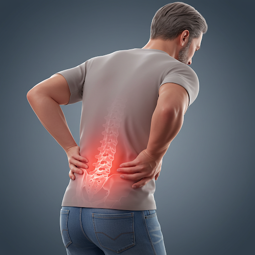
Manage Longstanding Low Back Pain by Spending Time in Nature
- At June 26, 2025
- By Healing In Motion
- In Research
 0
0
Study Overview
The article “Being away from everything”: Exploring the importance of access to nature for individuals living with chronic low back pain, published in the Journal of Pain, investigates how nature can aid in managing chronic low back pain (cLBP). It involved ten semi-structured interviews with people averaging 50.1 years old and living with cLBP for about 19.1 years.
Key Findings
The study found that nature can provide distraction, reduce isolation, and offer a calming environment, enhancing pain self-management. However, many face challenges like physical disabilities, environmental hazards, or lack of proximity to natural spaces. It suggests integrating nature-based activities into treatment plans and exploring virtual reality for those unable to access nature.
Detailed Analysis and Background
The article presents a pioneering qualitative cross-sectional study focused on understanding the role of natural environments in managing chronic low back pain (cLBP). The study, conducted and published in June 2025, addresses the growing evidence against the effectiveness of ‘one-size-fits-all’ pharmacological approaches, advocating for alternative non-pharmacological therapies such as green social prescribing and spending time in nature.
Methodology and Participants
The research methodology involved ten semi-structured interviews, primarily with women (9 out of 10 participants), with an average age of 50.1 years and an average duration of living with cLBP of 19.1 years. This qualitative approach aimed to explore the lived experiences of individuals, providing deep insights into their coping strategies and the role of nature in ameliorating pain. The analysis was conducted using Reflexive Thematic Analysis, identifying two main themes: (1) Importance of Nature and (2) Inaccessible Nature, each with four subthemes respectively.

Key Findings and Themes
The theme “Importance of Nature” underscores the benefits of accessing natural environments, which can significantly aid in self-management of cLBP. Participants reported that being in nature offered distraction, reduced feelings of isolation, and provided a calming environment, which helped alleviate pain and improve mental well-being. For instance, those able to immerse themselves in larger green spaces, such as forests, felt more positive, as they could lose themselves in the environment and focus less on their pain levels.
However, the theme “Inaccessible Nature” revealed significant barriers, including physical disabilities, environmental obstacles or hazards, and geographical limitations. These challenges often undermined the ability to access nature, highlighting an unmet clinical need. The study emphasizes that many individuals encounter substantial obstacles, such as uneven terrain, limited seating, or difficulties leaving their homes, making it challenging to benefit from the restorative properties of nature.
Implications and Recommendations
The findings suggest that integrating nature-based activities into treatment plans could be beneficial, offering a promising avenue for improving overall well-being for those with cLBP. The researchers recommend exploring virtual reality interventions to simulate nature experiences for those unable to access outdoor spaces, addressing accessibility issues. This approach could help make the benefits of nature accessible to everyone, potentially through technological innovations. Simple changes, such as better paths and seating, were also suggested to enhance real-world access.
The study marks the first qualitative investigation focusing directly on the nuanced role of natural settings within the coping mechanisms employed by people with cLBP. This pioneering aspect underscores the need for further research into how nature can be incorporated into holistic and multidisciplinary frameworks for treating pain, aligning with leading international organizations’ recommendations.
Contextual Background
Chronic low back pain is a major public health issue, with the highest prevalence globally among musculoskeletal conditions and being the leading cause of disability worldwide. It affects individuals of all ages, with peak cases at 50–55 years, and women experiencing it more frequently. The condition often leads to work loss, participation restriction, and reduced quality of life, making innovative approaches like nature-based interventions particularly relevant.
The study’s emphasis on accessibility challenges also aligns with broader discussions on health equity, addressing significant physical barriers faced by people living with chronic pain, as highlighted in the University of Plymouth news release. This includes the need for better infrastructure, such as improved paths and seating, to facilitate access to natural spaces.
Conclusion
In conclusion, the article provides valuable insights into the therapeutic potential of nature for individuals with cLBP, while also highlighting significant accessibility barriers. It advocates for innovative solutions like virtual reality to bridge these gaps, contributing to the broader discourse on holistic pain management. The findings are particularly timely, given the current understanding of cLBP as a major global health challenge, and offer a foundation for future research into nature-based interventions.

New Hope For Joint Repair
- At May 12, 2025
- By Healing In Motion
- In Research
- 0
A recent scientific breakthrough by Chen at al. could significantly improve how we treat cartilage damage — a common problem in joints like knees and hips that often leads to long-term pain and arthritis.
Cartilage is notoriously slow to heal, largely because its cells (called chondrocytes) have limited access to energy. To tackle this, researchers have developed a new technique that enhances the energy levels of these cells, helping them function better and regenerate damaged tissue more effectively.
The research team created energy-enhanced exosomes — tiny vesicles called Suc-EXO — which are loaded with high levels of ATP, the molecule that powers cell activity. When these Suc-EXO were delivered to stem cells and cartilage cells, they boosted energy metabolism and activated key repair pathways.
In particular, the treatment improved the transformation of stem cells into cartilage-producing cells and helped existing cartilage cells maintain their function by supporting healthy mitochondria (the cell’s energy centres).
Tests in a rabbit model with cartilage injuries showed that Suc-EXO, when delivered via a gel, significantly improved cartilage regeneration. The repaired tissue had higher levels of collagen type II and aggrecan — essential components of strong, healthy cartilage.
This innovative “organelle-tuning” approach opens up a new avenue for regenerative therapies and could one day lead to more effective treatments for joint injuries and osteoarthritis.
Chronic Low Back Pain, Bacterial Infection, and Antibiotic Treatment
- At April 21, 2025
- By Healing In Motion
- In Research
- 0
Chronic low back pain (CLBP) is a leading cause of disability globally, affecting millions and posing significant challenges to healthcare systems due to its complex aetiology and limited effective treatments (Gilligan et al., 2021). Recent research has sparked debate by suggesting that low-grade bacterial infections, particularly involving Cutibacterium acnes (C. acnes), may contribute to CLBP in a subset of patients, especially those with disc herniation and Modic Type 1 changes (bone oedema visible on MRI). This hypothesis, if validated, could revolutionise treatment by introducing antibiotics as an alternative to conventional therapies or invasive procedures like spinal surgery. However, the evidence remains controversial, with concerns about contamination, antibiotic efficacy, and stewardship. The following summary synthesises findings from key studies, highlighting microbiology evidence, clinical trial outcomes, and ongoing research challenges.
Microbiology Evidence: Bacterial Presence in Herniated Discs
Several studies have investigated whether bacteria, particularly C. acnes, are present in herniated disc tissue and contribute to CLBP. Gilligan et al. (2021) reviewed five well-designed microbiology studies that confirmed bacteria in disc samples, suggesting infection rather than contamination. These studies found C. acnes, a low-virulence anaerobic bacterium commonly associated with acne, in herniated disc tissue, particularly in patients with Modic Type 1 changes. However, the bacterial burden was low, potentially below detection limits in some studies, which may explain conflicting results where bacteria were absent or attributed to surgical contamination (Gilligan et al., 2021).
Astur et al. (2023) conducted a prospective cohort study to identify bacteria in herniated intervertebral discs, reporting C. acnes in a significant proportion of samples. Their stringent aseptic protocols minimised contamination risks, supporting the infection hypothesis. Similarly, Urquhart et al. (2015) conducted a systematic review and found moderate evidence that low-virulence bacteria are present in spinal disc material, particularly in patients with disc herniation and Modic Type 1 changes. They applied Bradford Hill’s criteria to assess causation, concluding modest evidence for a causal link but noting the need for further research to distinguish infection from contamination (Urquhart et al., 2015).
In contrast, Monge-García et al. (2024) explored whether bacterial presence in disc cultures represents true infection or contamination. Their study suggested that while C. acnes was frequently detected, contamination during surgical procedures could not be ruled out, particularly given the bacterium’s ubiquity on skin and in hair follicles. This raises a critical question: are bacteria in disc tissue causative agents of CLBP or incidental findings due to procedural artefacts? The conflicting findings underscore the need for standardised microbiological methods and larger sample sizes to clarify the role of C. acnes (Monge-García et al., 2024).
Clinical Evidence: Antibiotic Efficacy in CLBP
Clinical trials have tested whether antibiotics can reduce pain and disability in CLBP patients with suspected bacterial infections. Two randomised controlled trials (RCTs) reviewed by Gilligan et al. (2021) demonstrated significant pain and disability reductions in patients with CLBP and Modic Type 1 changes treated with oral antibiotics, typically amoxicillin-clavulanate, for up to 100 days. These patients, often unresponsive to conventional treatments like physiotherapy or analgesics, showed clinically meaningful improvements, suggesting antibiotics as a potential alternative to surgery for those facing disc replacement or fusion (Gilligan et al., 2021).
A landmark RCT by Albert et al. (2013), cited across multiple articles, found that 100 days of amoxicillin-clavulanate significantly reduced pain and disability in patients with CLBP post-disc herniation and Modic Type 1 changes compared to placebo. At one-year follow-up, the antibiotic group reported a median Roland Morris Disability Questionnaire (RMDQ) score reduction from 15 to 7, compared to 15 to 14 in the placebo group (p=0.0001). Pain scores and constant pain prevalence also improved significantly (Urquhart et al., 2015; Gilligan et al., 2021). However, the trial reported high rates of adverse events, particularly gastrointestinal issues like diarrhoea, in 65% of the antibiotic group versus 23% in the placebo group, raising concerns about tolerability (Urquhart et al., 2015).
Despite these promising results, not all studies support antibiotic efficacy. Some trials reported modest or non-significant effects, potentially due to underdosing or variable bacterial susceptibility. A review by Czaplewski et al. (2023) highlighted that oral amoxicillin doses (500–1000 mg, two or three times daily) may not achieve adequate intradiscal concentrations to target C. acnes effectively, with only 6.5% of serum concentrations reaching herniated disc tissue. Higher doses (e.g., 1000 mg three times daily) may be needed to reach efficacy targets for 90% of C. acnes strains, suggesting that previous studies may have been underdosed (Czaplewski et al., 2023).

Modic Changes and Infection Hypothesis
Modic Type 1 changes, characterised by vertebral bone oedema, are six times more prevalent in CLBP patients than the general population and are strongly associated with the infection hypothesis. Aebi (2013) noted that 80% of discs infected with C. acnes in surgical samples developed Modic Type 1 changes, suggesting that bacterial infection may trigger inflammation and oedema in adjacent vertebrae (Aebi, 2013). The neovascularisation following disc herniation is thought to allow C. acnes to enter the disc during transient bacteraemia, such as during tooth brushing, leading to chronic low-grade infection (Urquhart et al., 2015).
However, Modic changes may also result from mechanical stress or degeneration, complicating the attribution to infection alone. Lings (2013) and Rolfsen et al. (2024) emphasised that Modic Type 1 and Type 2 changes might represent different stages of a common degenerative process, questioning the specificity of infection as a cause. Rolfsen et al. (2024) proposed a multicentre case-control biopsy study to further investigate bacterial growth in CLBP patients with Modic changes, aiming to use advanced microbiological techniques like PCR to detect low-burden infections and control for contamination (Rolfsen et al., 2024).
Controversies and Challenges
The hypothesis that bacterial infection causes CLBP faces several challenges. First, the presence of C. acnes in disc tissue may reflect contamination rather than infection, given its prevalence on skin and difficulty in culturing due to its anaerobic nature (Monge-García et al., 2024). Second, the clinical significance of antibiotic benefits is debated, with some studies showing only modest effects and high adverse event rates (Gilligan et al., 2021). Third, long-term antibiotic use raises concerns about antimicrobial resistance and secondary infections like Clostridium difficile (Urquhart et al., 2015).
Antibiotic stewardship is a critical issue. The prolonged regimens (up to 100 days) used in trials contrast with typical short-course antibiotic treatments, increasing risks of resistance and side effects. Aebi (2013) and Lings (2013) cautioned against widespread antibiotic use without robust evidence, drawing parallels with the paradigm shift in peptic ulcer treatment following Helicobacter pylori discovery but urging caution until confirmatory studies are conducted.
Future Directions
The reviewed studies advocate for further research to refine patient selection, optimise antibiotic regimens, and address stewardship concerns. Rolfsen et al. (2024) and Czaplewski et al. (2023) emphasised the need for studies incorporating advanced diagnostics (e.g., quantitative microbiology, proteomics) and pharmacokinetic/pharmacodynamic evaluations to ensure adequate intradiscal antibiotic concentrations. Identifying biomarkers, such as cytokine patterns or genetic predispositions, could help select patients most likely to benefit from antibiotics (Gilligan et al., 2021).
Moreover, alternative delivery methods, such as intradiscal antibiotic injections (e.g., PP353, a novel treatment combining linezolid and a thermosensitive gel), are being explored to minimise systemic side effects and improve efficacy (Tripathi et al., 2025, cited in The Guardian). These approaches could offer targeted treatment for infection-related CLBP, reducing the need for prolonged oral antibiotics.
Conclusion
The hypothesis that low-grade bacterial infections contribute to CLBP, particularly in patients with disc herniation and Modic Type 1 changes, is supported by microbiological evidence and some clinical trials demonstrating antibiotic efficacy. However, controversies persist regarding contamination, variable clinical outcomes, and antibiotic stewardship. While antibiotics offer a potential alternative to surgery for a subset of CLBP patients, widespread adoption requires further validation through rigorous, multicentre studies. Future research should focus on optimising diagnostics, treatment regimens, and patient selection to balance efficacy with safety, potentially transforming the management of this debilitating condition.
Antibiotics For Low Back Pain?
- At March 31, 2025
- By Healing In Motion
- In Research
- 0
Persica Pharmaceuticals has developed PP353, an intradiscal injection designed to treat chronic low back pain linked to bacterial infections, particularly cases associated with Modic changes (pathological alterations in vertebrae seen on MRI). PP353 combines:
- Linezolid (antibiotic) – targets bacterial infection
- Iohexol (contrast agent) – aids imaging and distribution
- Thermosensitive gel – ensures controlled release
Recent Study & Findings
- Conducted on 44 patients
- Patients received two injections four days apart
- 60% reported significant pain and disability reduction
- Study supports infectious origins in some chronic back pain cases
Potential Impact
- Could benefit the 25% of chronic low back pain cases linked to bacterial infection
- Offers a minimally invasive alternative to surgery or long-term medication
- Further clinical trials required before widespread use
Cold Water Immersion and Health
- At February 20, 2025
- By Healing In Motion
- In Research
- 0
A recent systematic review and meta-analysis published in PLOS ONE aimed to evaluate the psychological, cognitive, and physiological effects of cold-water immersion (CWI) in healthy adults. The study synthesised data from 11 randomised controlled trials encompassing 3,177 participants who engaged in CWI through methods such as cold showers, ice baths, or plunges in water temperatures at or below 15°C for durations of at least 30 seconds.
Key Findings:
- Inflammation: The analysis revealed that CWI might reduce inflammation. This anti-inflammatory effect is particularly noted in athletic populations, where CWI is often utilised to mitigate exercise-induced muscle soreness. It is with noting that the anti-inflammatory effect may blunt the athlete’s adaption to exercise.
- Stress Reduction: While immediate reductions in stress levels were not observed post-immersion, a significant decrease in stress was reported 12 hours after CWI sessions. This delayed effect suggests that CWI may influence stress-related pathways over time, contributing to improved mental well-being.
- Immune Function: Regular exposure to cold showers was associated with a reduction in self-reported sickness absence from work. Participants who incorporated daily cold showers into their routines reported fewer instances of illness-related absences, indicating a potential enhancement of immune resilience.
- Quality of Life and Sleep: Short-term improvements in sleep quality and overall well-being were reported by participants following consistent CWI practices. However, these positive effects appeared to diminish after a period of 90 days, suggesting that the benefits of CWI on sleep and well-being may be transient and possibly require sustained or varied interventions to maintain.

Conclusion:
The study indicates that CWI may offer certain health benefits, particularly in stress reduction and immune function over time. However, the evidence is inconsistent, and many studies are limited by small sample sizes or a focus on athletic populations. Further comprehensive research is needed to fully understand the benefits and effects of cold-water immersion on health.
Processed Meats Increase Risk of Dementia
- At January 27, 2025
- By Healing In Motion
- In Research
- 0
A recent study published in Neurology on 11 February 2025, led by researchers from Brigham and Women’s Hospital, has investigated the relationship between long-term red meat consumption and the risk of dementia and cognitive decline in U.S. adults. The study utilised data from the Nurses’ Health Study and the Health Professionals Follow-Up Study, encompassing approximately 170,000 participants over a 43-year period.
Key Findings:
- Processed Red Meat and Dementia Risk: Individuals with the highest intake of processed red meats—such as bacon, sausages, and hot dogs—exhibited a 13% higher risk of developing dementia compared to those with lower consumption levels.
- Cognitive Decline: High consumption of processed red meats was associated with a 14% increase in subjective cognitive decline, indicating a perceived worsening of cognitive abilities over time.
- Unprocessed Red Meat: The study found that unprocessed red meats, such as beef, lamb, and pork, also contributed to cognitive risks, potentially due to harmful substances like nitrates, sodium, and saturated fats present in these meats.
Mechanisms Behind the Findings:
The detrimental effects of processed red meats on cognitive health are believed to be linked to several factors:
- Nitrates and Nitrites: Commonly used as preservatives in processed meats, these compounds can form nitrosamines, which have been implicated in neurodegenerative processes.
- High Sodium Content: Elevated sodium levels can lead to hypertension, a known risk factor for vascular dementia.
- Saturated Fats: High levels of saturated fats in processed meats are associated with cardiovascular diseases, which can indirectly affect brain health.

Dietary Recommendations:
Based on the findings, the researchers suggest several dietary modifications to mitigate dementia risk:
- Reduce Processed Red Meat Intake: Limiting the consumption of processed meats can lower the risk of cognitive decline.
- Substitute with Healthier Protein Sources: Replacing processed red meats with alternatives such as fish, poultry, nuts, and legumes has been associated with a reduced risk of dementia. For instance, substituting processed red meat with fish was linked to a 28% reduction in dementia risk.
- Adopt Brain-Healthy Diets: Diets like the Mediterranean, DASH, and MIND diets, which are low in red meat and rich in fruits, vegetables, whole grains, and healthy fats, have been associated with decreased dementia risks.
Conclusion:
This extensive study underscores the importance of dietary choices in maintaining cognitive health. Limiting the intake of processed red meats and opting for healthier protein sources may significantly reduce the risk of dementia and support long-term cognitive function.
Food Choices Affect Pain
- At December 31, 2024
- By Healing In Motion
- In Research
- 0
Introduction
Chronic pain is a widespread issue that significantly impacts quality of life and healthcare systems. While obesity is a well-documented risk factor for chronic pain, recent evidence suggests that other factors, such as diet quality, may also play a role. A recent study explores the relationship between diet quality and body pain in adults, regardless of their levels of adiposity (body fat).
Objective
The primary aim was to investigate whether better diet quality is linked to lower levels of body pain in adults and whether this association remains significant independent of adiposity.
Methods
The research used data from the Whyalla Intergenerational Study of Health, which includes a diverse sample of adults from Whyalla, South Australia. Participants completed detailed assessments, including:
- Diet quality evaluation: Using the Australian Recommended Food Score (ARFS), which measures adherence to dietary guidelines.
- Body pain measurement: Using validated self-reported questionnaires that assess the severity and frequency of pain across the body.
- Adiposity indicators: These included body mass index (BMI), waist circumference, and body fat percentage.
The study also controlled for confounding factors such as age, sex, physical activity, smoking status, and socio-economic status.
Key Findings
- Diet Quality and Pain:
- Higher ARFS scores, indicating better diet quality, were significantly associated with reduced levels of body pain.
- The association was consistent across various demographic and lifestyle groups.
- Independence from Adiposity:
- While higher adiposity was associated with increased body pain, the relationship between diet quality and pain remained significant even after adjusting for adiposity measures.
- This suggests that diet quality has an independent role in pain modulation.
- Potential Mechanisms:
- Nutritional components such as anti-inflammatory foods, antioxidants, and omega-3 fatty acids might contribute to reduced systemic inflammation and pain perception.
- Conversely, poor diet quality may exacerbate pain through increased inflammation and metabolic dysregulation.
Implications
The findings highlight the importance of a high-quality diet in managing body pain, regardless of body fat levels. This has potential implications for public health strategies and clinical interventions aimed at reducing chronic pain prevalence.
- For individuals: Encouraging adherence to dietary guidelines may help alleviate body pain alongside other health benefits.
- For policymakers and healthcare providers: Integrating dietary advice into pain management protocols could improve patient outcomes.
Conclusion
The study concludes that better diet quality is associated with reduced body pain in adults, and this relationship is independent of adiposity. These results underscore the importance of dietary quality as a modifiable factor in chronic pain prevention and management. Further research is needed to explore specific dietary components and their mechanisms in influencing pain pathways.
This research supports the broader understanding that nutrition plays a critical role in overall health and highlights diet quality as a key factor in pain management strategies.
Changes in Gait, Balance and Strength with Aging
- At November 3, 2024
- By Healing In Motion
- In Research
- 0
The recent article “Age-related changes in gait, balance, and strength parameters: A cross-sectional study” published in PLOS One by Rezaei et al. examines the impact of aging on various physical performance measures such as gait (walking patterns), balance, and muscular strength. The study employs a cross-sectional design, meaning it analyses data collected at one specific point in time across a diverse sample of adults ranging in age, with the objective of understanding how these physical parameters change as people grow older.
Background and Motivation
The motivation behind this study stems from the known correlation between age-related declines in physical abilities and increased risk of falls, decreased independence, and overall diminished quality of life. As gait, balance, and strength are crucial to maintaining mobility and preventing falls, identifying when and how these factors decline can inform interventions that help older adults retain their independence longer. The study also aims to provide insights for clinicians and healthcare professionals to tailor preventative measures and rehabilitation strategies according to age-specific needs.
Methods
The researchers recruited a large group of participants spanning various age groups and used a range of objective measurements to assess gait, balance, and strength. For gait analysis, the study evaluated variables such as walking speed, stride length, and step time variability. Balance was measured through static and dynamic assessments, which included tests for single-leg standing time and balance stability during movement. Strength was assessed primarily through grip strength and lower limb muscle power tests, which are widely accepted indicators of general muscular strength in ageing populations.
Findings
The study found significant age-related declines across all parameters, with notable differences between age groups:
- Gait: There was a clear trend of reduced walking speed and shorter stride length with increasing age, coupled with an increase in gait variability. These changes often started to manifest in middle-aged adults and progressively worsened in older age groups.
- Balance: Balance deficits were observed as early as middle age, with a marked reduction in single-leg standing time and stability during dynamic movements in older adults. The results highlight that both static and dynamic balance abilities diminish with age, increasing the risk of falls.
- Strength: Muscle strength, particularly grip strength and lower limb power, also showed a steady decline with age. This decline was particularly significant in participants over 60 years, indicating that muscle weakness becomes more prominent and impactful in later years.

Interpretation and Implications
The findings suggest that the aging process is associated with measurable declines in gait, balance, and strength, which collectively heighten the risk of falls and mobility impairments. The study highlights that these changes do not occur suddenly but rather develop gradually, implying that early interventions in middle-aged adults could be beneficial. The researchers suggest that routine assessment of gait, balance, and strength could be integrated into clinical practice to detect early declines and support proactive management strategies.
Conclusion
In summary, this cross-sectional study provides valuable insights into how age impacts physical performance parameters crucial for mobility and independence. The age-related deterioration in gait, balance, and strength underlines the importance of early assessment and targeted exercise or rehabilitation interventions aimed at mitigating these declines. By identifying the specific onset of these changes, healthcare providers can better design preventative programs tailored to the needs of ageing adults, thereby potentially enhancing quality of life and reducing the social and economic burdens associated with falls and functional impairments in the elderly population.
This study reinforces the significance of personalised health strategies and preventive measures in addressing the age-related decline in physical function, advocating for policies that encourage physical activity and strength maintenance from a younger age.
Rugby, Concussion and Neurodegenerative Disease
- At October 7, 2024
- By Healing In Motion
- In Research
- 0
The article “Concussion-Related Biomarker Variations in Retired Rugby Players and Implications for Neurodegenerative Disease Risk: The UK Rugby Health Study“ delves into the long-term health impacts of concussions on retired rugby players, particularly focusing on the risk of neurodegenerative diseases like Alzheimer’s, Parkinson’s, and chronic traumatic encephalopathy (CTE). The study aims to explore the relationship between repeated head injuries and variations in specific biomarkers associated with brain health, which may indicate increased susceptibility to these diseases.

Key areas of focus include:
- Biomarker Identification:
The study identifies several biomarkers in the blood and cerebrospinal fluid (CSF) that are linked to brain trauma. These biomarkers include proteins like tau and neurofilament light (NFL), both of which are known to rise following concussions and other brain injuries. The analysis of these markers in retired rugby players is crucial in detecting early signs of brain damage. - Concussion History and Biomarker Variations:
A significant finding from the study is that retired players with a history of multiple concussions showed elevated levels of these biomarkers compared to those with fewer or no reported concussions. This correlation suggests that repeated head impacts may lead to lasting changes in brain structure and function, potentially increasing the risk of cognitive decline or neurodegenerative disease. - Implications for Neurodegenerative Disease Risk:
The elevated levels of biomarkers such as tau and NFL in retired players may serve as early indicators of brain degeneration, which is often associated with conditions like CTE. The study draws attention to the possibility that these players are at a higher risk of developing neurodegenerative disorders compared to the general population, emphasising the importance of long-term health monitoring. - Preventive and Diagnostic Recommendations:
Based on the findings, the study calls for better concussion management strategies during athletes’ active careers, particularly in high-contact sports like rugby. Regular monitoring of retired players for biomarker changes could also help in early detection and intervention, potentially mitigating the risk of serious brain diseases. This includes the use of advanced imaging techniques and blood tests to track changes over time. - Broader Impact on Contact Sports:
The study contributes to the broader understanding of concussion-related brain injuries in contact sports, reinforcing concerns about the long-term health of athletes who experience repeated head trauma. It also raises ethical and medical considerations for rugby and other sports in how they manage and address player health both during and after their careers.
In conclusion, the research highlights a pressing need to reassess how concussions are treated in rugby, not just for immediate recovery, but for long-term brain health. By focusing on biomarker variations, the study offers a new avenue for detecting early signs of neurodegeneration, potentially helping to reduce the incidence of conditions like CTE among retired rugby players.
The Musculoskeletal Syndrome of Menopause
- At September 8, 2024
- By Healing In Motion
- In Research
- 0
Wright el al. have recently published a review article on the musculoskeletal syndrome of menopause – a complex issue that affects millions of women worldwide.
The Role of Estrogen in Musculoskeletal Health
Estrogen plays a crucial role in maintaining bone density, muscle mass and tendon structure. It helps to:
- Stimulate bone formation: Estrogen promotes the activity of osteoblasts, cells responsible for building new bone tissue.
- Inhibit bone resorption: It reduces the activity of osteoclasts, cells that break down bone tissue.
- Maintain muscle mass: Estrogen helps to regulate muscle protein synthesis and breakdown, preventing excessive muscle loss.
- Maintain tendon structure and function: It influences collagen metabolism and maintains optimal tendon stiffness.
- Maintain healthy joints: Estrogen helps decrease inflammation and maintain joint structure.
When estrogen levels decline during menopause, these processes become imbalanced, leading to bone loss, increased risk of fractures, muscle atrophy, joint pain and stiffness, and increased predisposition to tendinopathies and risk of tendon ruptures.

Risk Factors for Musculoskeletal Syndrome of Menopause
Several factors can increase a woman’s risk of developing musculoskeletal issues during or after menopause:
- Genetics: A family history of osteoporosis or other bone disorders can increase susceptibility.
- Lifestyle factors: Smoking, excessive alcohol consumption, and a sedentary lifestyle can contribute to bone loss and muscle weakness.
- Nutrition: Inadequate intake of calcium and vitamin D can impair bone health.
- Medical conditions: Certain conditions, such as autoimmune diseases or thyroid disorders, can affect bone metabolism.
Treatment and Prevention Strategies
While there is no cure for the musculoskeletal syndrome of menopause, effective management strategies can help alleviate symptoms and improve quality of life. These include:
- Hormone replacement therapy (HRT): When used appropriately, HRT can help maintain bone density and reduce the risk of fractures. However, it’s important to discuss the potential risks and benefits with a healthcare provider.
- Lifestyle modifications: Regular exercise, especially weight-bearing activities, can help strengthen bones and muscles. A balanced diet rich in calcium, vitamin D, and protein is also crucial.
- Medications: In some cases, medications like bisphosphonates or selective estrogen receptor modulators (SERMs) may be prescribed to treat osteoporosis.
- Supplements: Calcium and vitamin D supplements can be beneficial for maintaining bone health.
By understanding the causes, risks, and treatment options for the musculoskeletal syndrome of menopause, women can take proactive steps to protect their health and well-being during this important life stage.